Parkinson's triangle

Synonyms
Triangular Space of Parkinson, Infratrochlear triangle

The Parkinson triangle, also known as the Triangular Space of Parkinson, was first described in 1965 by D. Parkinson as an approach to the Cavernous sinus[^1].

Anatomy

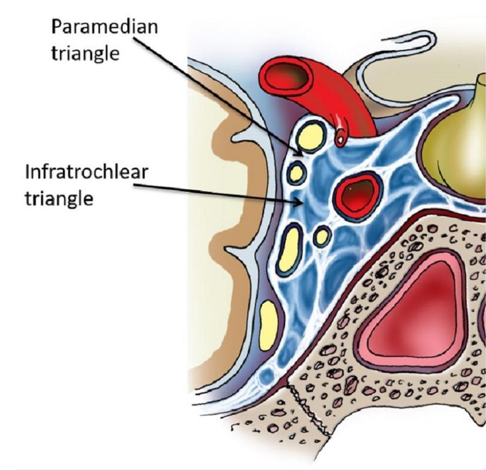
The Parkinson triangle is formed by the first division of the Trigeminal nerve (Ophthalmic nerve), the Trochlear nerve, and the lateral wall of the Cavernous sinus[^2].

Figure

Parkinson triangle
Visualization of the infratrochlear triangle (Parkinson triangle) and the paramedian triangle in the region of the cavernous sinus. Figure adapted from Drazin, Doniel, et al. Intracranial anatomical triangles: a comprehensive illustrated review. Cureus 9.10 (2017).

Editorial transparency

Authorship & revisions

Who created and updated this article, and which changes are documented.

Article created by
Last edited:
on
Revision history
4 edits · 1 contributor
Author list:
Show revision history 4 entries

4 edits by 1 author

Edit · Alaric Steinmetz ·

No content change recorded

Edit · Alaric Steinmetz · (+126 −0)

No content change recorded

Edit · Alaric Steinmetz · (+0 −135)

No content change recorded

Article created on · Alaric Steinmetz

Image viewer

Image

Open original