Internal cerebral vein
Last edit by Alaric Steinmetz on
Synonyms: Internal cerebral vein
ICD-11: XA6DC3
The internal cerebral vein is a paired deep cerebral vein that drains into the great cerebral vein.
Anatomy
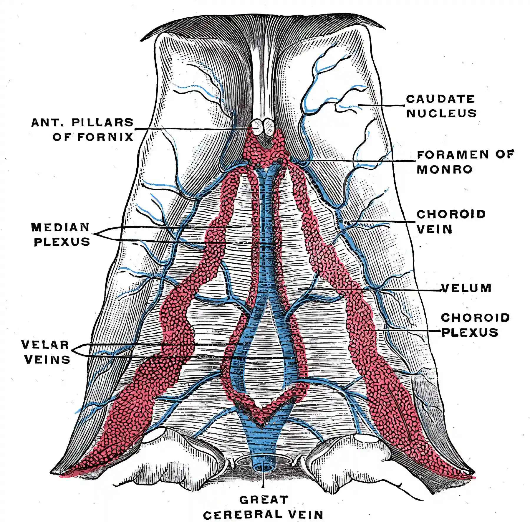
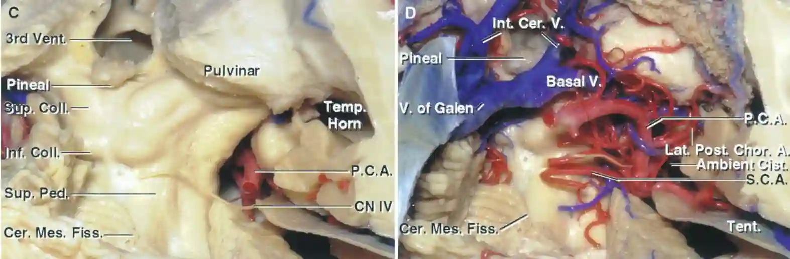
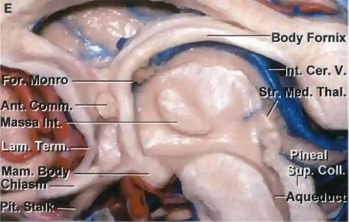
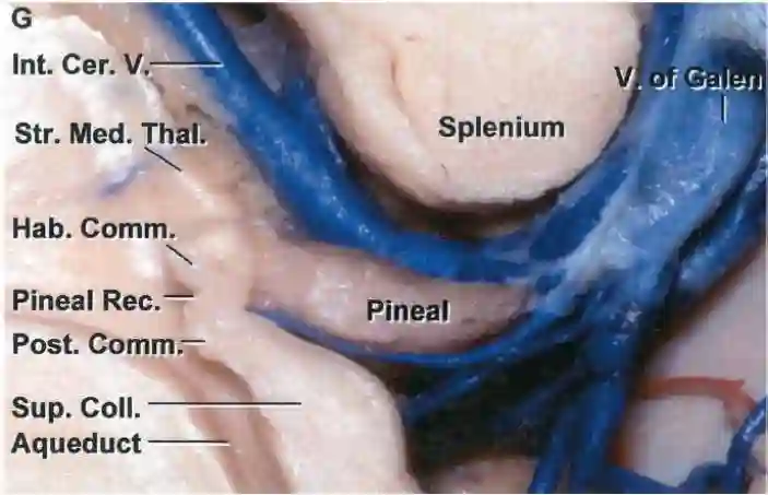
The internal cerebral vein originates from multiple venous tributaries in the region of the foramen of Monro and runs posteriorly in the roof of the third ventricle above the striae medullaris thalami between the two layers of the tela choroidea. The anterior parts of the internal cerebral vein run parallel to each other adjacent to the midline. Posteriorly, they diverge to run along the superolateral surface of the pineal gland. Further posteriorly, below the splenium of the corpus callosum, the two internal cerebral veins converge and, after leaving the velum interpositum in the quadrigeminal cistern, flow into the great cerebral vein[^1].
Clinical Relevance
An occlusion of the internal cerebral vein (e.g. due to thrombosis or surgically) can have severe clinical consequences and lead to venous infarctions[^2].
Illustration


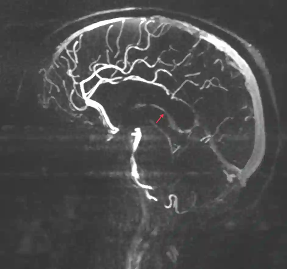
Imaging