The internal acoustic meatus runs within the Temporal bone and is part of the inner ear.
Anatomy
Overall, the internal acoustic meatus extends approximately 1 cm within the temporal bone. The internal acoustic meatus opens via the internal acoustic porus into the posterior cranial fossa. The following anatomically relevant structures pass through the internal acoustic meatus:
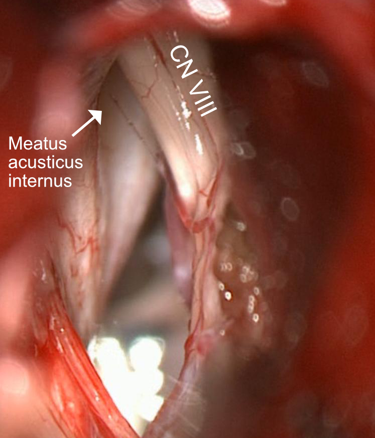
EnlargeSchematic illustration of the anatomical structures in the internal auditory canal, showing the crista transversa. Figure adapted from A. Rhoton.EnlargeDepiction of the vestibulocochlear nerve (CN VIII) and the internal acoustic meatus during a retrosigmoid approach. The facial nerve is obscured by the vestibulocochlear nerve and is not visible.
Imaging
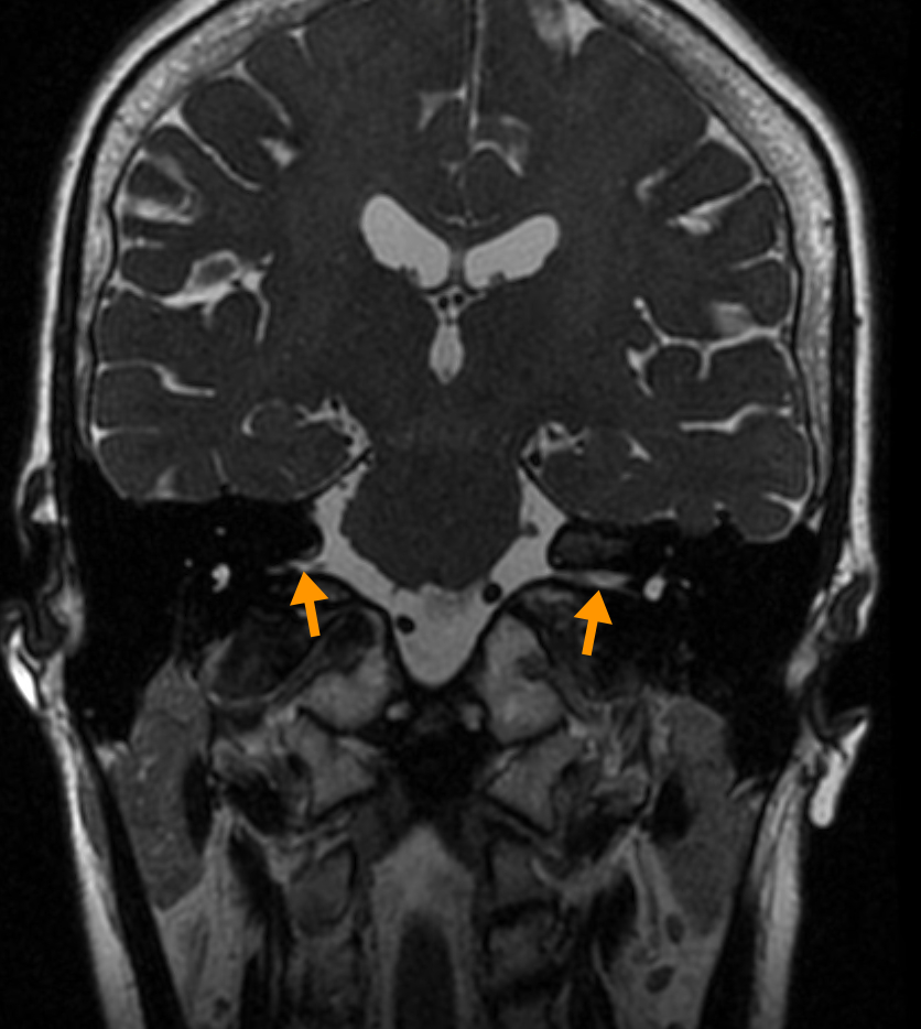
EnlargeVisualization of the internal auditory canal (red arrow) on an axial CT image.EnlargeDepiction of the internal acoustic meatus (orange arrow) on a coronal T2-weighted MRI image of the neurocranium.
Weekly update
Newsletter
Relevant neurosurgical studies, concisely summarized in a weekly email newsletter.
By subscribing, you consent to receiving the newsletter through Buttondown. Unsubscribe using the link in any email.
Privacy policy