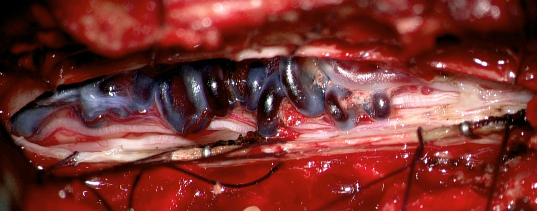
Dural arteriovenous fistulas (dAVFs) are acquired vascular malformations that arise at the level of the dura mater (Dura mater). They are direct arteriovenous shunts.
Epidemiology
Intracranial dAVFs
Dural arteriovenous fistulas account for approximately 10 % to 15 % of intracranial vascular malformations and are slightly overrepresented in the posterior cranial fossa, at approximately 35 %. The annual incidence of intracranial dAVFs is approximately 0.16 \/ 100000.
Spinal dAVFs
The annual incidence of spinal dural arteriovenous fistulas is approximately 0.5 \/ 100000.
Classification
Cranial dural arteriovenous fistulas are usually classified according to the Cognard or Borden classification.
Imaging
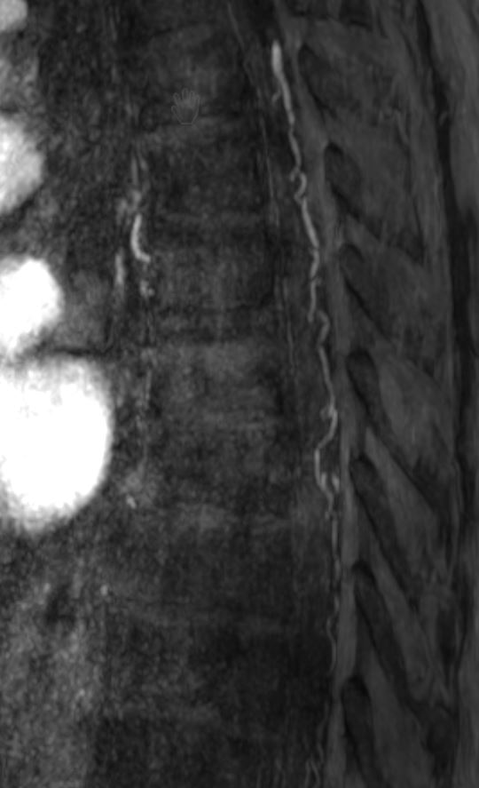
The gold standard for evaluating dural arteriovenous fistulas is Digital subtraction angiography. MR angiography also allows dural arteriovenous fistulas to be visualized.
Risk of hemorrhage
The risk of hemorrhage correlates with the presence of retrograde cortical venous drainage. Overall morbidity is 15 % and mortality is 10 % in the presence of retrograde cortical venous drainage[^2]. In a meta-analysis of 377 cases[^4], three dAVF locations with a particularly aggressive course were identified (Aggressive:Benign ratio):
Tentorial (31:1)
Middle cranial fossa\/Sylvian (2.5:1)
Anterior cranial fossa\/ethmoidal (2.1:1)
Treatment
Treatment of dural arteriovenous fistulas aims to eliminate the risk of hemorrhage and progressive neurological deficits by occluding the dAVF at the fistulous point. dAVFs in the anterior cranial fossa always drain[^3] via cortical veins and therefore require a more aggressive treatment approach[^1].