EnlargeVisualization of Dorello canal. Figure adapted from Umansky et al.EnlargeVisualization of the ligamentum sphenopetrosum (labeled Petrosph. Lig.), which forms the cranial boundary of Dorello’s canal. Figure adapted from Yasuda A. et al. 2004.
Imaging
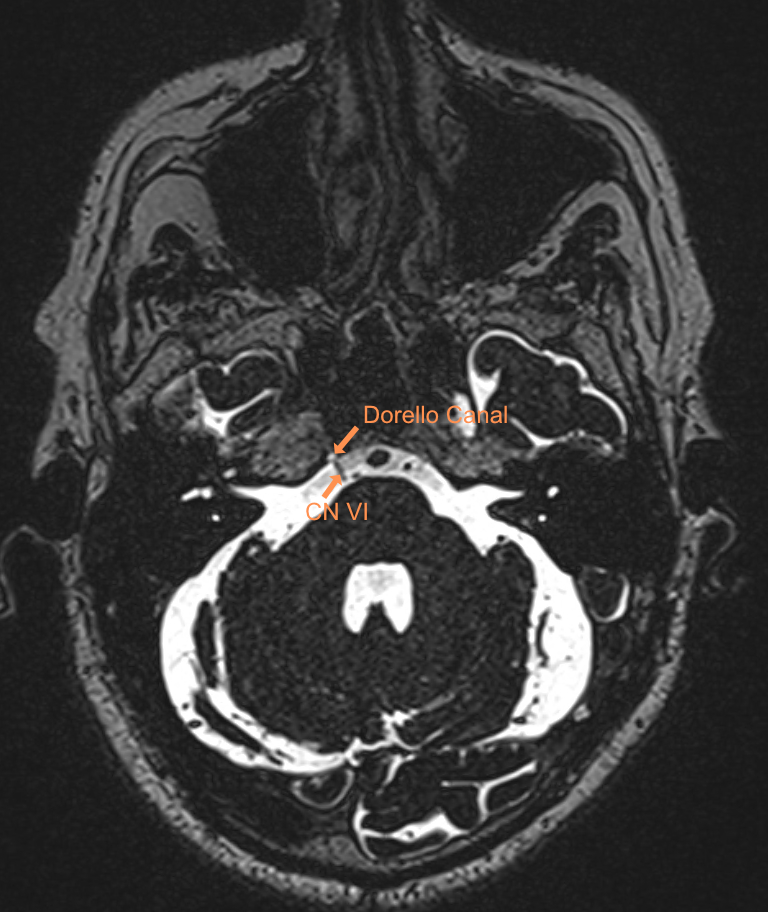
EnlargeVisualization of the abducens nerve (CN VI) and Dorello canal on axial T2 imaging.
Weekly update
Newsletter
Relevant neurosurgical studies, concisely summarized in a weekly email newsletter.
By subscribing, you consent to receiving the newsletter through Buttondown. Unsubscribe using the link in any email.
Privacy policy