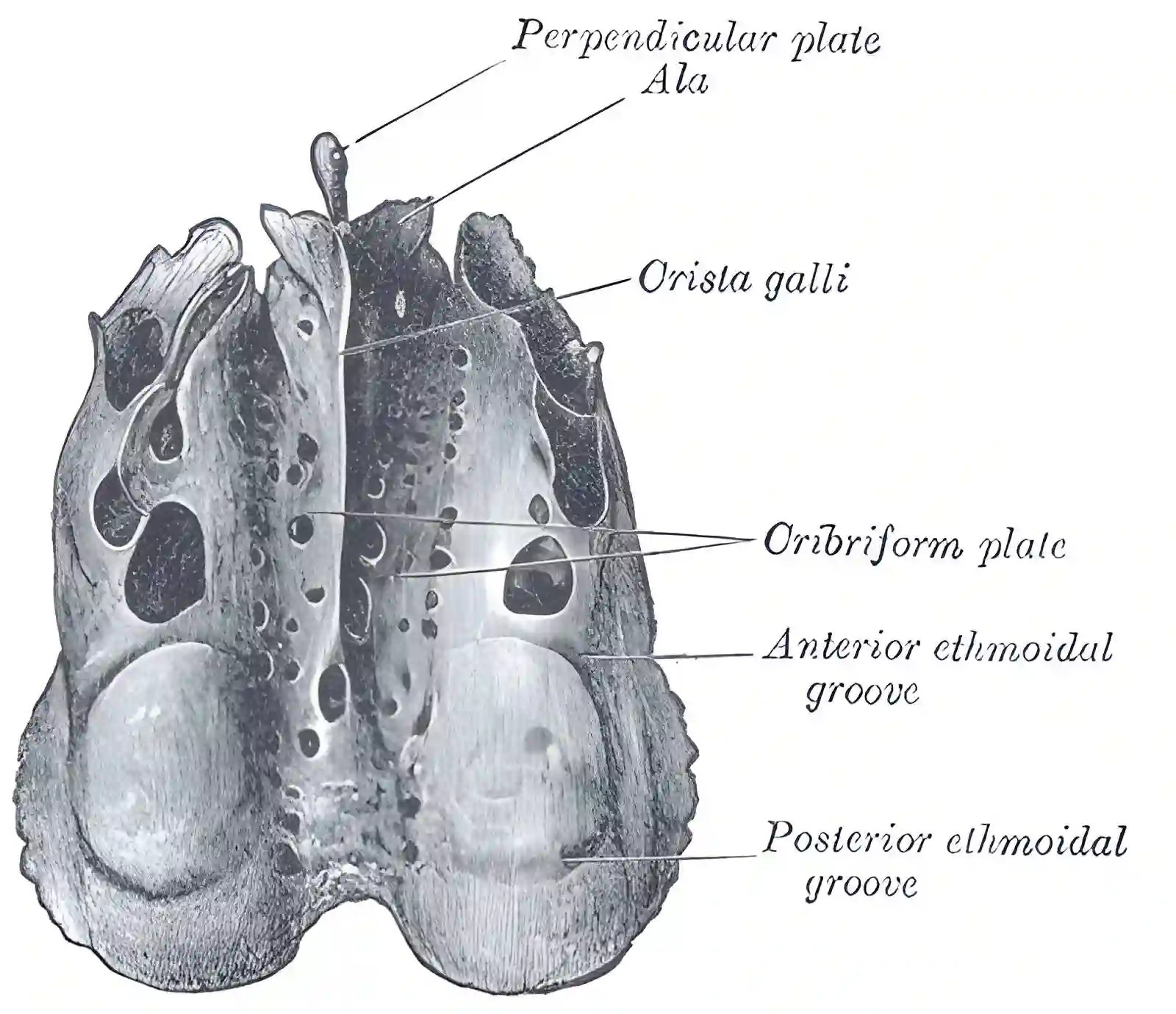
Crista galli
Last edit by Alaric Steinmetz on
The Crista galli is a bony projection of the ethmoid bone. At the posterior edge of the crista galli, the falx cerebri attaches.
Embryology
Embryologically, the cista galli develops from the ethmoid bone. In the second fetal month, the crista galli forms together with other structures of the anterior cranial fossa. Ossification of the crista galli begins approximately 2 months postnatally and is completed after about 24 months[^1].
Pneumatization
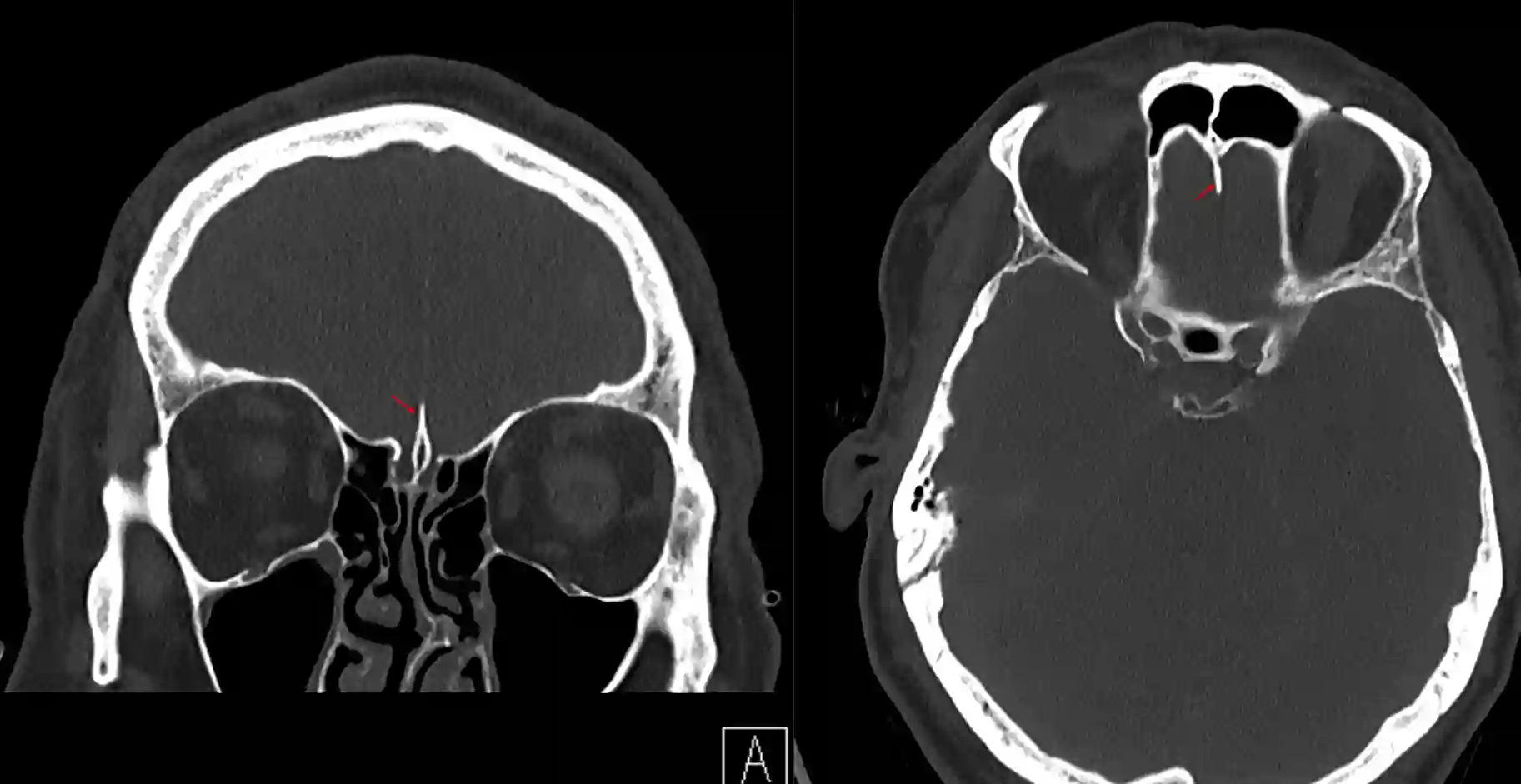
The crista galli is morphologically variable depending on the individual and can be pneumatized. Pneumatization of the crista galli is primarily an extension from the frontal sinus[^2].
Illustration


Imaging