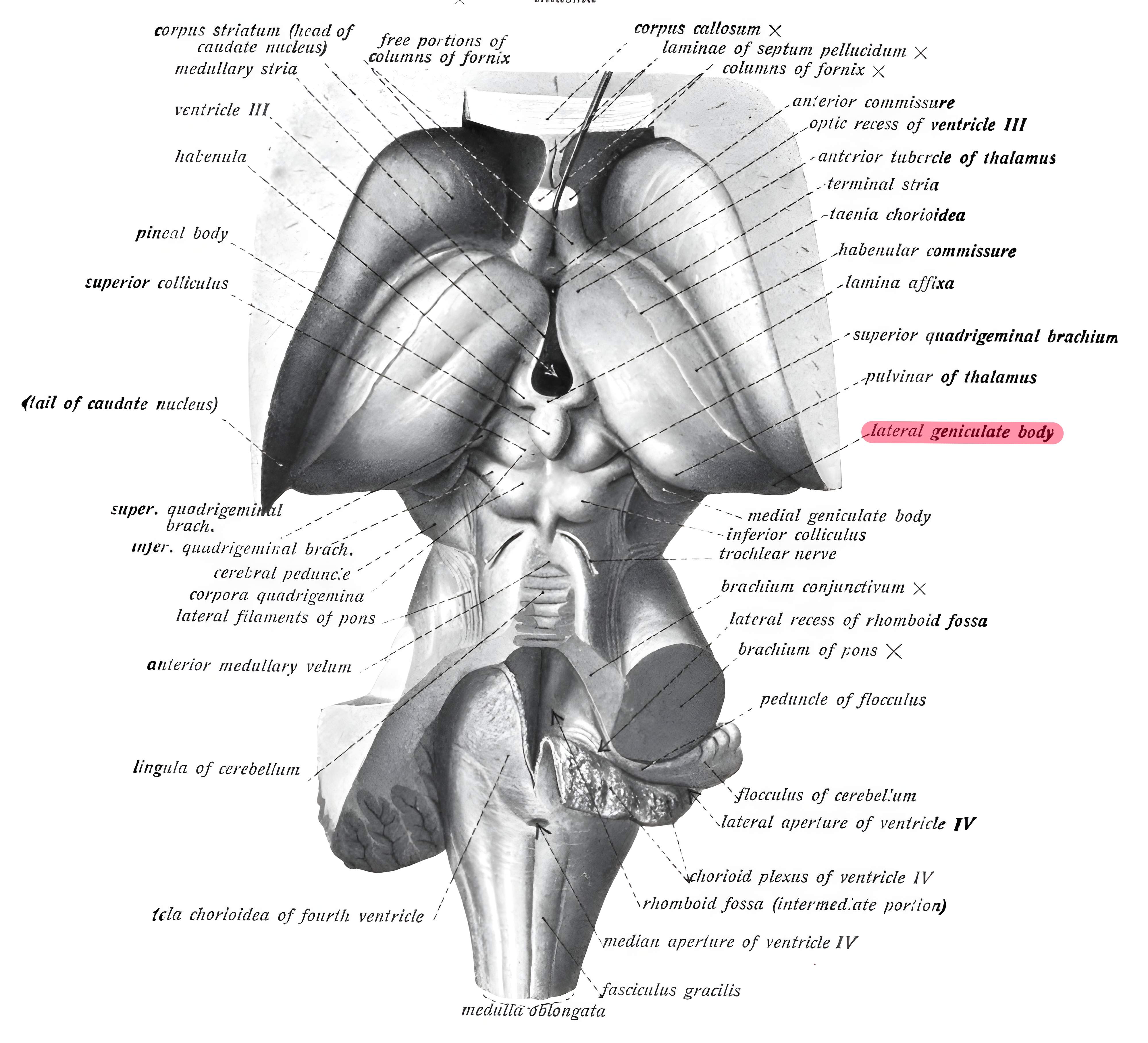
The corpus geniculatum laterale is a nuclear region in the Diencephalon and part of the visual pathway.
Anatomy
The corpus geniculatum laterale is located on the ventrocaudal surface of the Thalamus and constitutes a relatively independent structure. The corpus geniculatum laterale is organized into six layers of cells, which are separated by the incoming fiber bundles of the optic tract.
Afferents
In each of the two corpora geniculata, crossed and uncrossed optic nerve fibers terminate. The left corpus geniculatum laterale represents the temporal half of the retina of the left eye and the nasal half of the retina of the right eye, while the right corpus geniculatum laterale represents the temporal half of the retina of the right eye and the nasal half of the retina of the left eye. Fibers from the macula, the region of sharpest vision, terminate in a central wedge-shaped region that extends through all cell layers.
Efferents
The efferents of the corpus geniculatum laterale travel as the central visual radiation, the radiatio optica, to the visual cortex, the striate area on the medial hemispheric surface of the occipital lobe[^1].