Cavum vergae
Last edit by Alaric Steinmetz on
Synonyms: CV
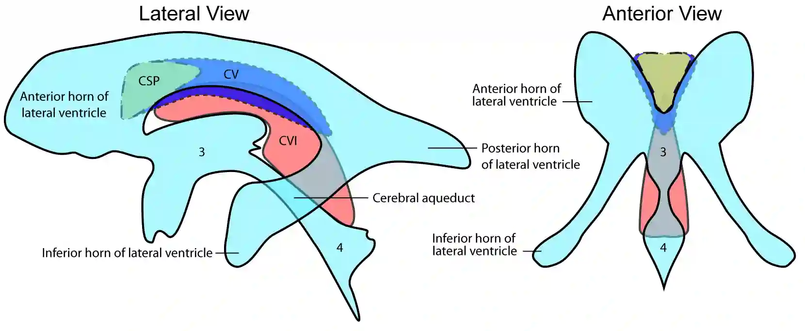
The cavum vergae is referred to as a cerebrospinal fluid-filled duplication of the septum pellucidum dorsal to the foramen interventriculare.
Anatomy
A cavum vergae often occurs together with the cavum septi pellucidi and corresponds to a dorsal extension of the latter.
Clinical Relevance
The cavum vergae is an anatomical norm variant and is usually not clinically noticeable, typically identified as an incidental finding in imaging. In the literature, associations with developmental delays, macrocephaly, and Apert's syndrome are described[^1].
Illustration

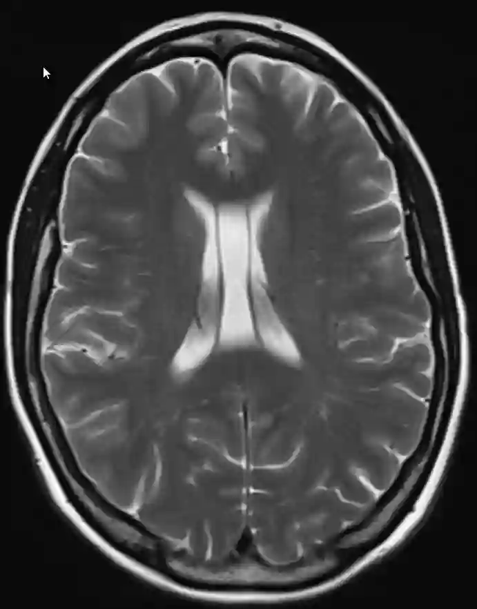
Imaging