An aspergilloma is a fungal infection caused by molds of the genus Aspergillus. Aspergillomas most commonly occur in the lungs, but they can also affect the central nervous system (CNS), where they represent a rare but life-threatening disease. Aspergillomas occur predominantly in immunocompromised individuals, but can also affect immunocompetent patients.
Pathophysiology
Aspergillus spores are ubiquitous in the environment and are inhaled by humans on a daily basis. In individuals with a healthy immune system, these spores are generally eliminated without difficulty. However, in the setting of impaired immune defenses, as occurs in patients after organ transplantation, in leukemia, or during long-term corticosteroid therapy, the spores can germinate and invade tissue. The most common route by which Aspergillus reaches the CNS is hematogenous dissemination from a primary site of infection, usually the lungs. Less commonly, spread occurs directly (per continuitatem) from adjacent structures such as the paranasal sinuses or the Petrous bone. In rare cases, direct inoculation of spores may also occur after neurosurgical procedures or cranial trauma. Once in the brain, the fungus tends to invade blood vessels, which can lead to thromboses, infarctions, and Hemorrhages. The resulting lesion is typically an encapsulated Abscess, the aspergilloma, consisting of a dense network of fungal hyphae, inflammatory cells, and necrotic tissue.
Symptoms
The clinical presentation of an intracranial aspergilloma is nonspecific and depends on the location and size of the lesion. Common symptoms include headache, fever, focal neurological deficits, seizures, and disturbances of consciousness.
Diagnosis
A definitive diagnosis requires histopathological identification of fungal hyphae in a tissue sample, which is generally obtained by biopsy or during resection. In addition, cultures can be performed on the collected material to identify the specific Aspergillus species and enable targeted antifungal therapy. Detection of Aspergillus antigens (e.g., galactomannan) in the blood or Cerebrospinal fluid may also be suggestive, but is not always reliable[^1].
Imaging
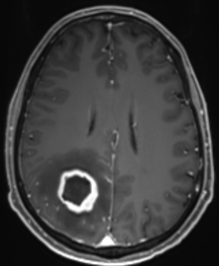
MRI is the gold standard for imaging aspergillomas, which appear radiographically like an abscess.
Treatment
The gold standard in the treatment of cerebral aspergilloma is surgical resection. The goal of the procedure is to remove the fungal mass and abscess capsule as completely as possible. This has several advantages:
Diagnostic confirmation: Obtaining tissue for histopathological and microbiological examination.
Mass reduction: Decompression of the surrounding brain tissue and reduction of intracranial pressure.
Improved drug efficacy: Removal of the necrotic mass improves the penetration of antifungal agents into the remaining infected tissue.
Simple aspiration of the encapsulated aspergilloma contents frequently results in recurrence and necessitates an additional open surgical procedure[^2].