Arachnoid cysts are benign, congenital cystic abnormalities of the arachnoid mater. They are usually asymptomatic cysts and therefore only rarely require surgical treatment. The presence of an arachnoid cyst may increase the risk of intracranial hemorrhage1. Rarely, arachnoid cysts may show progressive enlargement over time and thereby cause symptoms due to mass effect2.
Epidemiology
Arachnoid cysts occur in approximately 1 % of the population3.
Symptoms
Arachnoid cysts are typically asymptomatic. In a case series of 20 patients who underwent surgery for an arachnoid cyst, the following symptoms were observed in symptomatic patients4:
| Symptom | Frequency n (%) |
|---|---|
| Headache | 9 (41 %) |
| Weakness | 5 (23 %) |
| Seizure | 3 (14 %) |
| Visual loss | 1 (4 %) |
| Cognitive decline | 1 (4 %) |
| Hydrocephalus | 2 (9 %) |
| Scoliosis | 1 (4 %) |
Location
Arachnoid cysts may occur at various intracranial locations as well as spinally5 and are distributed by frequency as shown in the following list:
| Location | Frequency in %6 |
|---|---|
| Sylvian fissure | 49 |
| Cerebellopontine angle | 11 |
| Supracollicular | 10 |
| Vermis | 9 |
| Sellar & suprasellar | 9 |
| Interhemispheric | 5 |
| Cerebral convexity | 4 |
| Clivus | 3 |
Classification
Arachnoid cysts located in the middle cranial fossa can be classified into three types according to the Galassi classification.
Surgical treatment
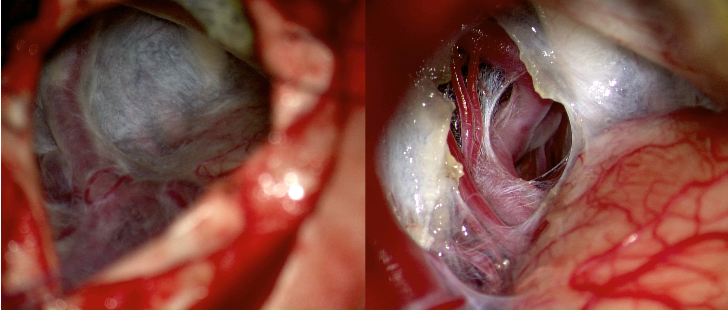
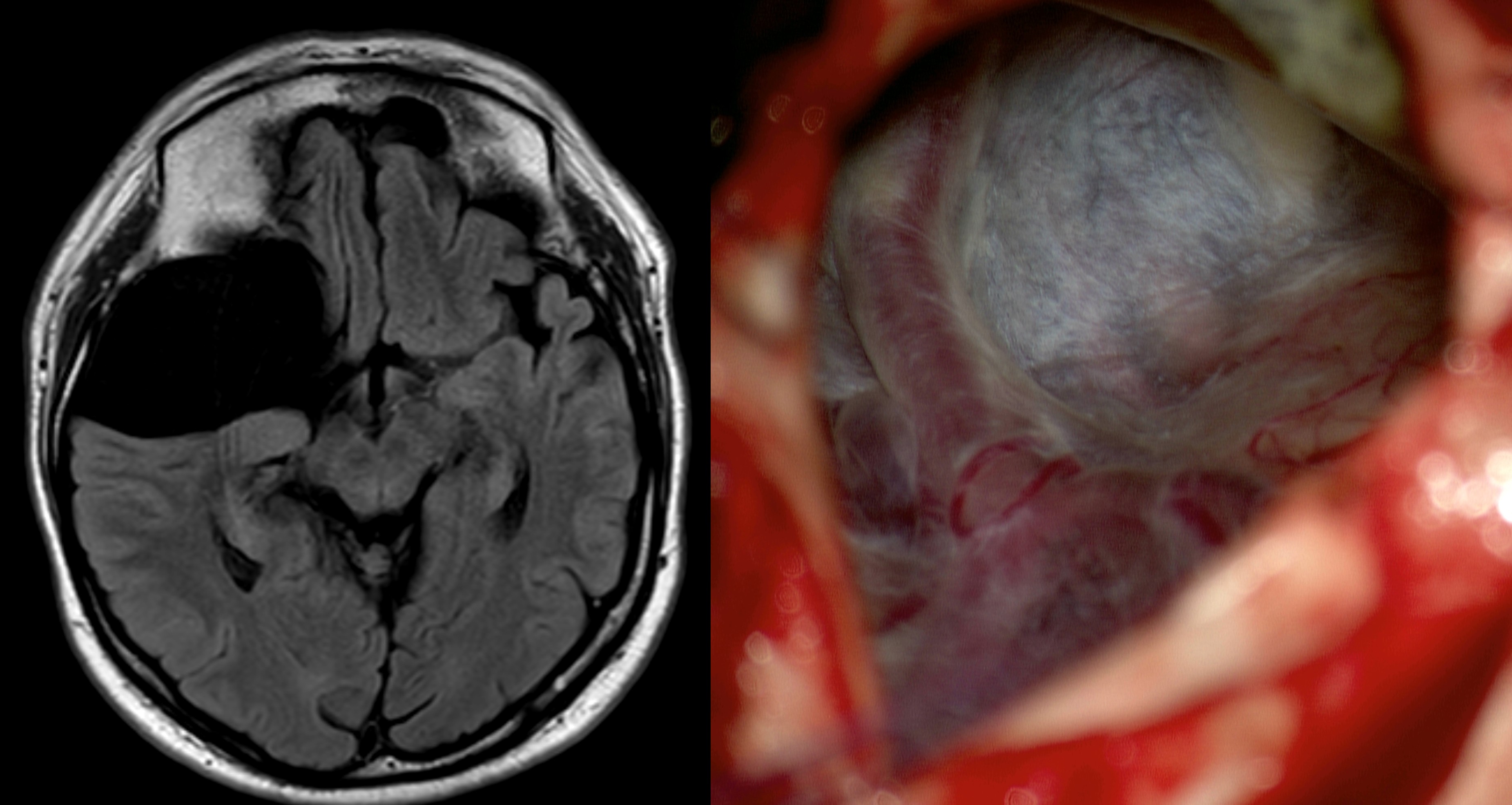
The majority of patients with arachnoid cysts do not require treatment. In rare cases, surgical intervention may be indicated for symptomatic arachnoid cysts. Typically, surgical fenestration of the arachnoid cyst is performed.
Complications
In a case series of 20 patients who underwent surgery for an arachnoid cyst, the following postoperative complications were observed7:
| Complication | Frequency (n) |
|---|---|
| None | 11 |
| Weakness | 3 |
| Cyst reaccumulation | 5 |
| Need for shunt placement | 3 |
| CSF fistula | 1 |
| Seizure | 1 |
| Subdural hygroma | 1 |
Intraoperative
Imaging
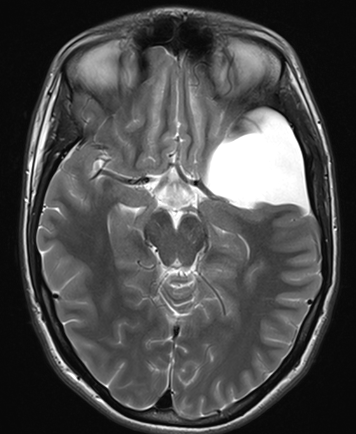
Arachnoid cysts can be visualized on CT as well as MRI.


References
Book: Neurosurgical Knowledge Update - A comprehensive review. Robert E. Harbaugh et al. Thieme Verlag. ↩ ↩ ↩ ↩ ↩ ↩
Pradilla, Gustavo, and George Jallo. "Arachnoid cysts: case series and review of the literature." Neurosurgical focus 22.2 (2007): 1-4. ↩ ↩ ↩ ↩ ↩ ↩
Rengachary SS, Watanabe I. Ultrastructure and Pathogenesis of Intracranial Arachnoid Cysts. J Neuropathol Exp Neurol. 1981;40:61-83. ↩ ↩