Subthalamic nucleus
Last edit by Alaric Steinmetz on
Synonyms: STN, Subthalamic nucleus, Corpus Luysii
The subthalamic nucleus (STN) is functionally part of the Basal ganglia and plays an important role in extrapyramidal motor function.
Anatomy
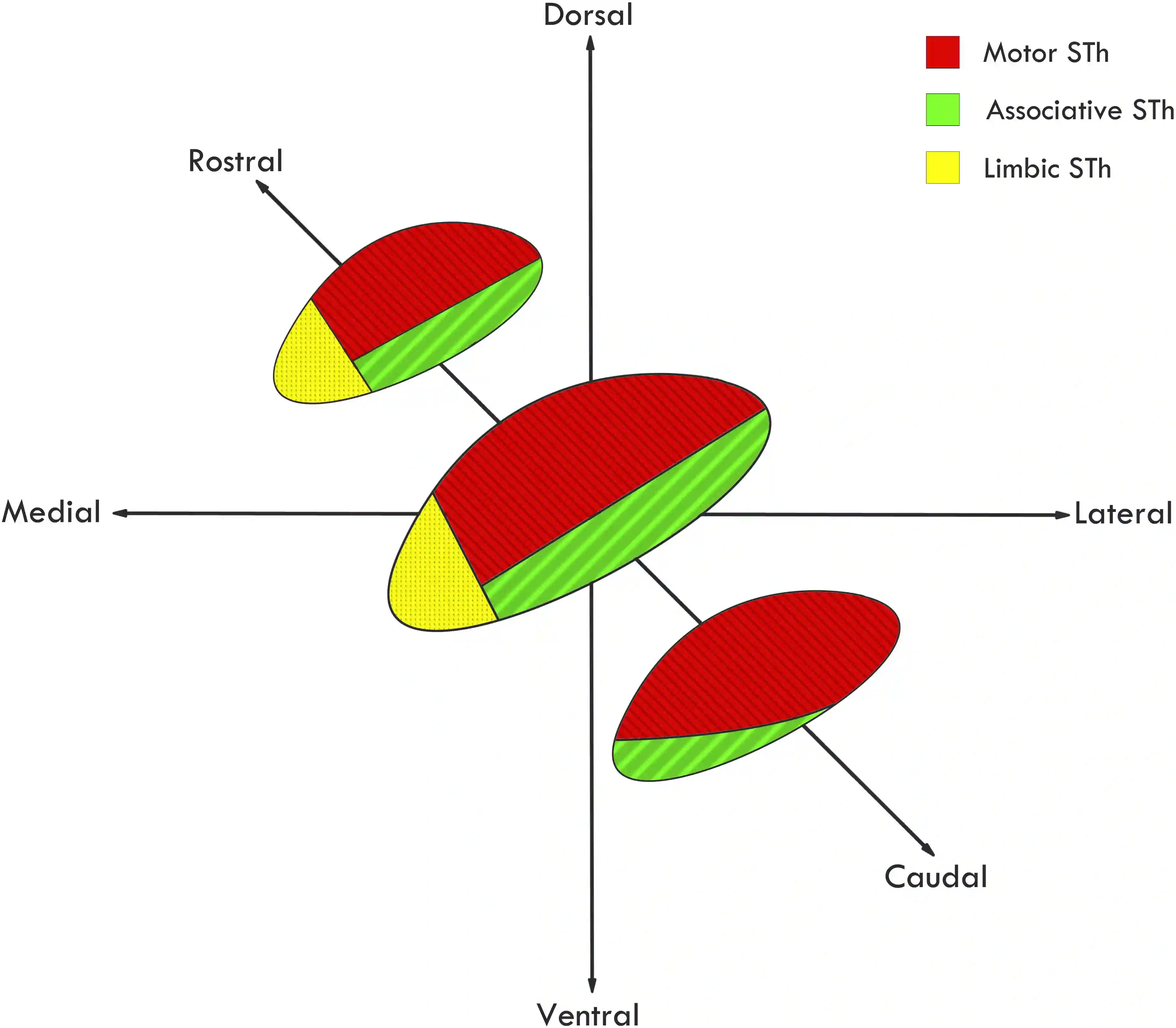
The subthalamic nucleus is oval in shape and is located ventral to the Thalamus in the Diencephalon; it can be functionally divided into limbic, associative, and motor subdivisions [^4].

Imaging
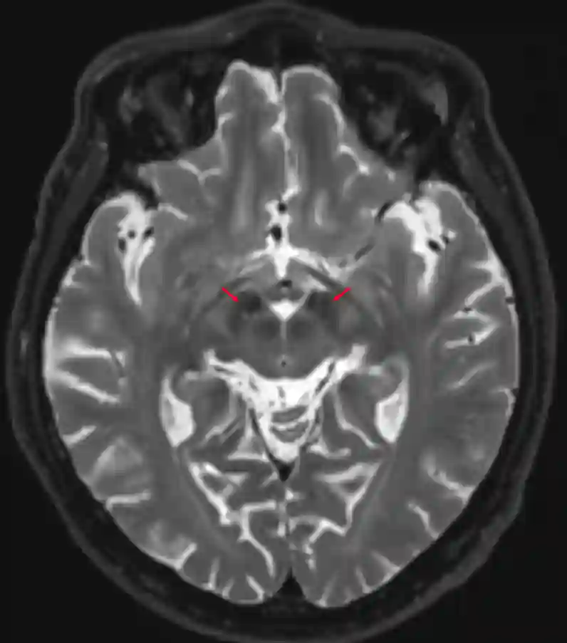
The subthalamic nucleus can be visualized particularly well on MRI using a T2, SWI, or FLAIR sequence [^2].

Clinical relevance
In patients with Parkinson's disease who qualify for Deep Brain Stimulation (DBS), the subthalamic nucleus is one of the possible target structures for electrode implantation. Stimulation of the subthalamic nucleus can particularly improve the motor symptoms of Parkinson's disease, whereas the nonmotor symptoms remain largely unaffected [^1].
Lesions
Occlusion of the perforating branches of the posterior communicating artery can lead to impairment of the subthalamic nucleus [^3]. A lesion in the subthalamic nucleus can result in the clinical presentation of Hemiballismus.