The pineal gland is also known as the epiphysis or pineal gland.
Anatomy
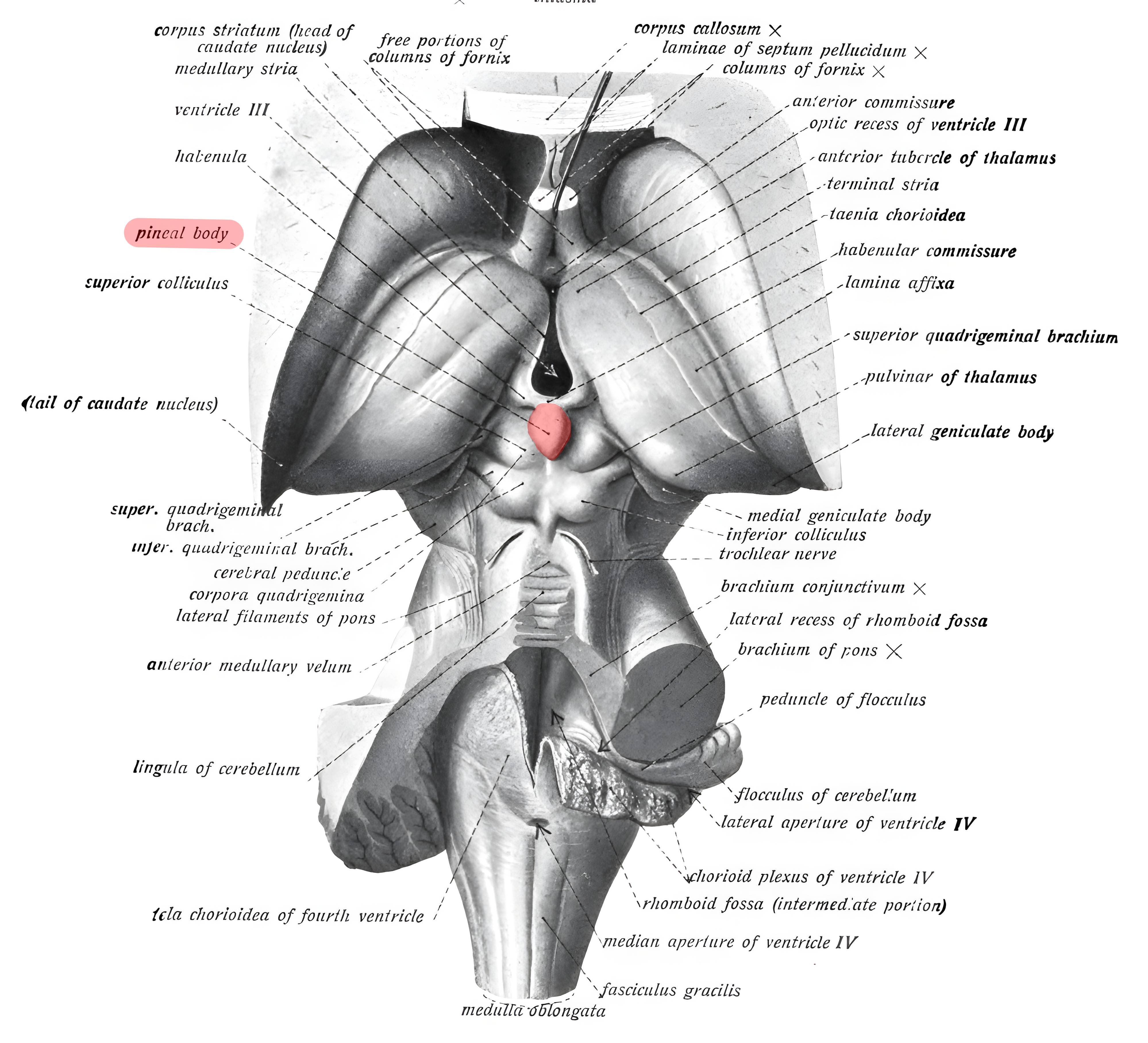
The pineal gland is located as part of the Epithalamus on the posterior wall of the third ventricle above the quadrigeminal plate.
Histology
The pineal gland is surrounded by a connective tissue capsule. The neuroendocrine cells of the pineal gland, the pinealocytes, are divided into lobules by connective tissue septa.
Hormones
Pinealocytes synthesize and secrete indoles and peptides, particularly α-melanocyte-stimulating hormone (α-MSH) and melatonin[^1].
Figure
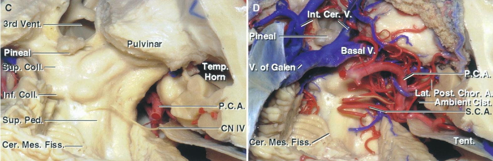
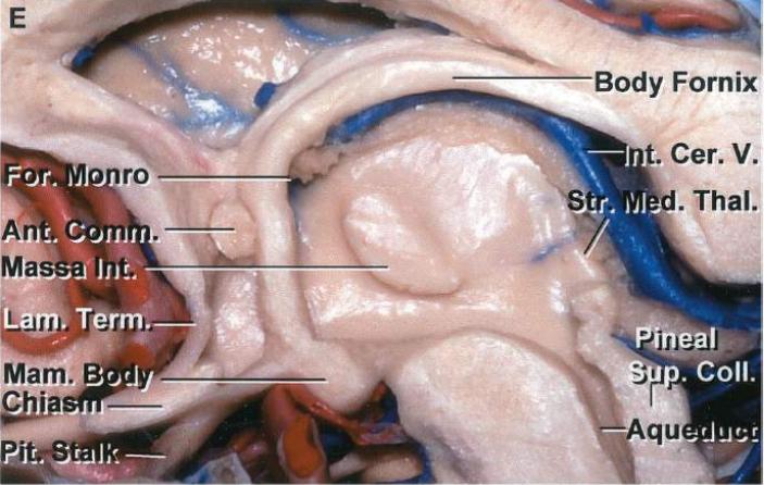
EnlargeSchematic depiction of the pineal gland (colored red) on an anatomical drawing from Sobotta's Atlas and Textbook of Human Anatomy, 1908 (Figure 650).EnlargeAnatomical illustration of the quadrigeminal plate with and without the corresponding vascular structures. Illustration adapted from Albert Rhoton 2002 Neurosurgery.EnlargeSagittal section through the third ventricle. Figure adapted from Albert Rhoton 2002 in Neurosurgery.
Imaging
EnlargeVisualization of the pineal gland on a sagittal T1-weighted sequence from a 7 Tesla MRI.
Weekly update
Newsletter
Relevant neurosurgical studies, concisely summarized in a weekly email newsletter.
By subscribing, you consent to receiving the newsletter through Buttondown. Unsubscribe using the link in any email.
Privacy policy