Mandibular nerve
Last edit by Alaric Steinmetz on
Synonyms: Mandibularnerv
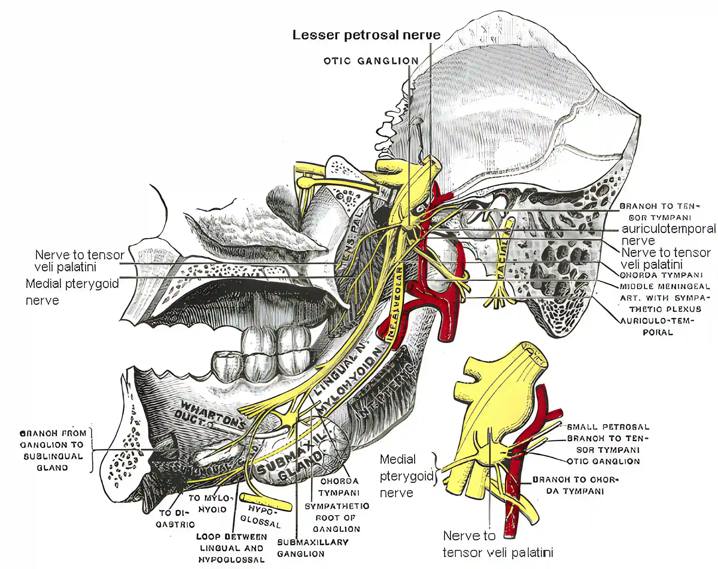
The mandibular nerve is the third (V3) and largest branch of the trigeminal nerve.
Anatomy
The mandibular nerve carries both sensory and motor fibers and exits through the foramen ovale from the base of the skull into the infratemporal fossa. After exiting, it divides into several branches that supply various regions of the face, oral cavity, and masticatory muscles. The main branches include the auriculotemporal nerve, the lingual nerve and the inferior alveolar nerve[^1].
Function
The mandibular nerve has a dual function: it is both sensory and motor. Sensory fibers transmit touch, pain, and temperature sensations from the lower jaw, teeth, gums, anterior two-thirds of the tongue, and parts of the face. Motor fibers control the masticatory muscles, which are responsible for chewing and the movement of the lower jaw. Additionally, it innervates the mylohyoid muscle and the anterior belly of the digastric muscle. The lingual nerve, a sensory branch, also plays a role in taste sensation as it carries fibers from the chorda tympani (a branch of the facial nerve) that relay taste signals from the tongue. The auriculotemporal nerve has an additional function in the parasympathetic innervation of the parotid gland by transmitting fibers from the otic ganglion.
Illustration