Homer-Wright Rosette

Homer-Wright rosettes can be observed in various tumors. They consist of a rosette-shaped arrangement of cells with a central lumen containing a meshwork of fiber-rich neuropil[^1].

Figure
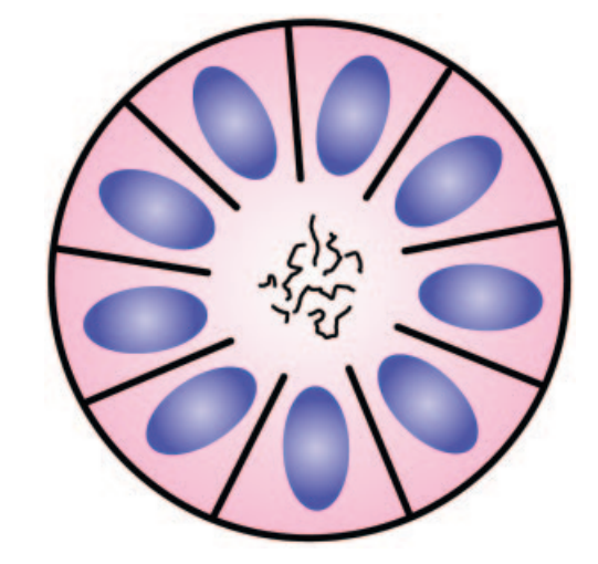
Homer-Wright rosette
Schematic illustration of a Homer-Wright rosette.
Histopathology of Homer-Wright rosettes
Multiple Homer-Wright rosettes in a patient with a PNET. H&E staining at ×400 magnification. Figure adapted from Wippold et al. 2006.

Associated Tumors

The presence of Homer-Wright rosettes is associated with the following tumors[^1]:

Editorial transparency

Authorship & revisions

Who created and updated this article, and which changes are documented.

Article created by
Last edited:
on
Revision history
5 edits · 1 contributor
Author list:
Show revision history 5 entries

5 edits by 1 author

Edit · Alaric Steinmetz ·

No content change recorded

Edit · Alaric Steinmetz · (+113 −0)

No content change recorded

Edit · Alaric Steinmetz · (+0 −5)

No content change recorded

Edit · Alaric Steinmetz ·

No content change recorded

Article created on · Alaric Steinmetz

Image viewer

Image

Open original