Foramen spinosum

The foramen spinosum is a small bony opening at the base of the skull

Anatomy

The foramen spinosum is located in the sphenoid bone and serves as a passage for the middle meningeal artery. The middle meningeal vein and the recurrent meningeal branch of the mandibular nerve also pass through it1. In humans, the foramen spinosum can be observed as early as 8 months after birth2.

Figure
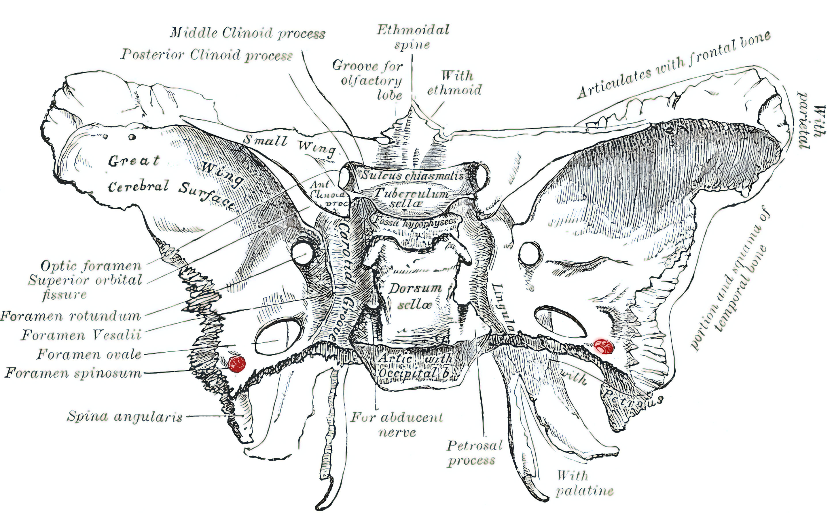
Foramen spinosum
Schematic illustration of the foramen spinosum (marked in red) in the sphenoid bone. Figure adapted from Gray's Anatomy.

Imaging

Foramen spinosum on CT
Foramen spinosum in three different planes using a bone-window CT image.

References


    1. Krayenbühl, Niklaus, Gustavo Rassier Isolan, and Ossama Al-Mefty. "The foramen spinosum: a landmark in middle fossa surgery." Neurosurgical review 31.4 (2008): 397. ↩ ↩

    2. Yanagi, S. "Developmental studies on the foramen rotundum, foramen ovale and foramen spinosum of the human sphenoid bone." The Hokkaido journal of medical science 62.3 (1987): 485-496. ↩ ↩

    Editorial transparency

    Authorship & revisions

    Who created and updated this article, and which changes are documented.

    Article created by
    Last edited:
    on
    Revision history
    8 edits · 1 contributor
    Author list:
    Show revision history 8 entries

    8 edits by 1 author

    Edit · Alaric Steinmetz · (+2 −2)

    No content change recorded

    Edit · Alaric Steinmetz ·

    No content change recorded

    Edit · Alaric Steinmetz · (+38 −0)

    No content change recorded

    Edit · Alaric Steinmetz · (+0 −36)

    No content change recorded

    Edit · Alaric Steinmetz ·

    No content change recorded

    Edit · Alaric Steinmetz · (+38 −0)

    No content change recorded

    Edit · Alaric Steinmetz · (+0 −36)

    No content change recorded

    Article created on · Alaric Steinmetz

    Image viewer

    Image

    Open original