Flow Void Signal

Last edit by Alaric Steinmetz on

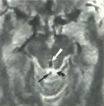
The phenomenon of a Flow Void Signal can be observed in MRI imaging and manifests as a signal loss on MRI due to the flow velocity of fluids, such as Cerebrospinal fluid or blood[^1].

Example

For example, in Aqueductal stenosis, a loss of the Flow Void Signal occurs due to the absence of cerebrospinal fluid flow.

Figure

Flow void signal on MRI
Axial T2 MRI image. A flow void signal is seen in the aqueduct (white arrow). The CSF in the cisterns is hyperintense (curved black arrow). Blood flow in the vein (straight black arrow) also appears dark with a flow void signal. Figure adapted from Sherman et al.

Article information

Article created by
Last edited:
on
Revision history
Show revision history (3)

3 edits by 1 author

Edit · Alaric Steinmetz · (+95 −0)

No content change recorded

Edit · Alaric Steinmetz · (+0 −91)

No content change recorded

Article created on · Alaric Steinmetz

Author list:
Newsletter