Corpus callosum
Last edit by Alaric Steinmetz on
The corpus callosum is a transverse fiber connection between the two cerebral hemispheres and forms the largest part of the commissural system.
Anatomy
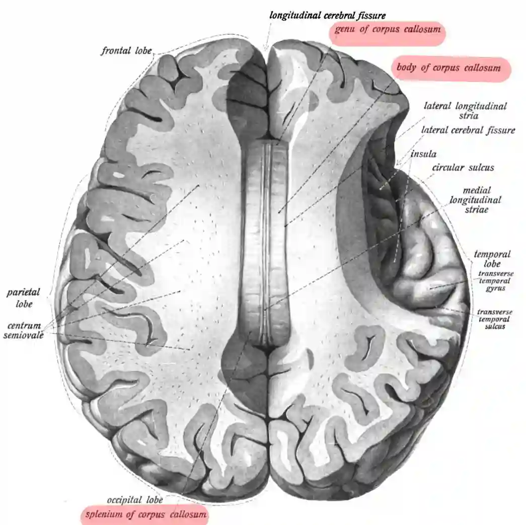
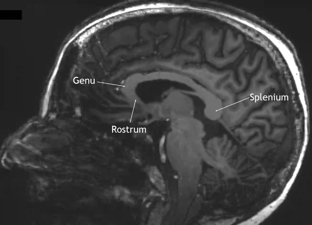
The corpus callosum is anatomically divided into three parts: rostrum, genu, and splenium.
Rostrum corporis callosi
The rostrum corporis callosi refers to the anterior, tapering section of the corpus callosum beneath the genu corporis callosi. It is connected to the anterior commissure and the lamina terminalis.
Genu corporis callosi
The genu corporis callosi is the anterior part of the corpus callosum facing the two frontal lobes, which tapers into the rostrum corporis callosi.
Splenium corporis callosi
The splenium corporis callosi describes the posterior thickened section of the corpus callosum.
Figure

Imaging

Clinical relevance
Common lesions that manifest in the corpus callosum are lymphomas, multiple sclerosis plaques, lipomas, or diffuse axonal injuries after trauma[^1].