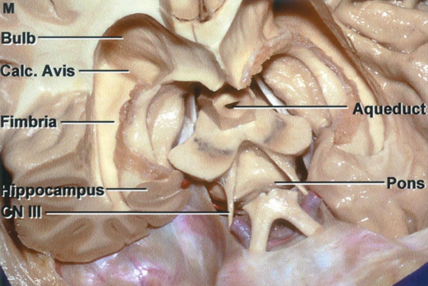
The calcar avis is a protrusion in the medial wall of the posterior horn of the lateral ventricle.
Anatomy
The calcar avis is caused by the deeply invaginating Calcarine sulcus. The calcar avis is prominent in approximately 24 % of cases, with a deep sulcus calcarinus[^1].
Figure
Clinical relevance
The calcar avis may be visualized on ultrasound examinations, for example through the fontanelles, or intraoperatively. On ultrasound, the calcar avis may be misinterpreted as a blood clot or mass lesion[^2].