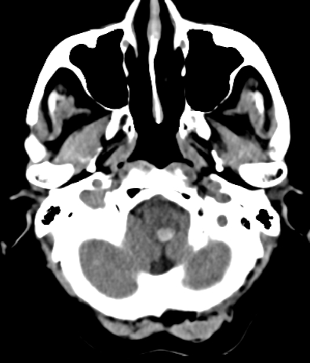
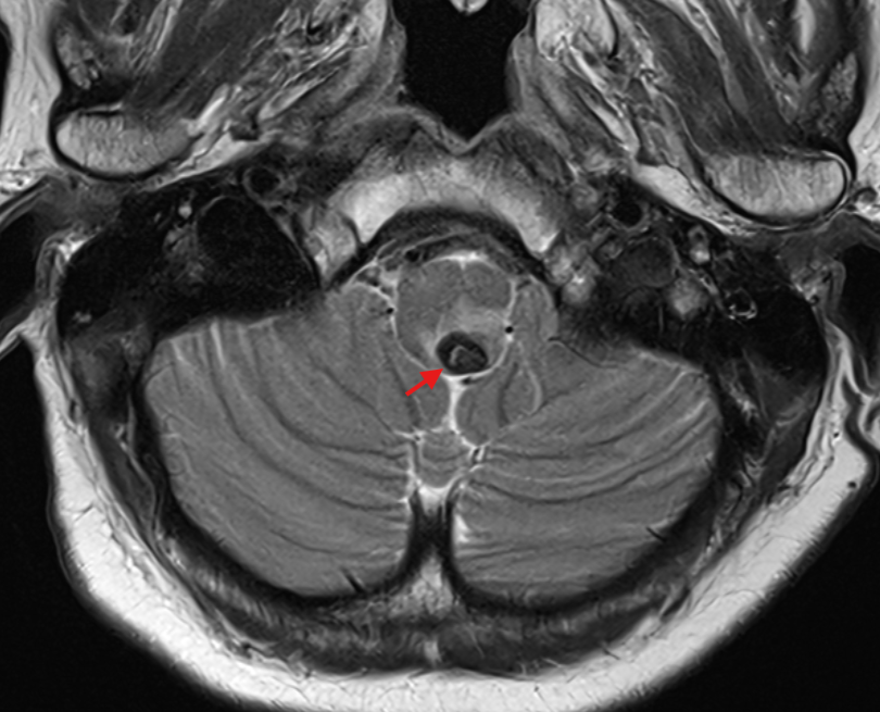
A brainstem cavernoma is a cavernous malformation located in the Brainstem.
Risk of hemorrhage
Cavernomas in this location have an annual risk of hemorrhage of 4 to 5 % per lesion per year[^1].
Imaging
MRI is the gold standard imaging modality for diagnosing brainstem cavernomas.
EnlargeMRI of a brainstem cavernoma in the left dorsolateral medulla oblongata on a sagittal MRI image.EnlargeAxial T2-weighted MRI sequence of a patient with a cavernoma in the left dorsolateral medulla oblongata.EnlargeAxiale Darstellung eines Hirnstammkavernoms in einer CT-Bildgebung.EnlargeDarstellung eines Hirnstammkavernoms (roter Pfeil) in einer axialen T2 Sequenz im MRT.
Weekly update
Newsletter
Relevant neurosurgical studies, concisely summarized in a weekly email newsletter.
By subscribing, you consent to receiving the newsletter through Buttondown. Unsubscribe using the link in any email.
Privacy policy