Glandula pinealis

Last edit by Alaric Steinmetz on

Synonyms: Epiphyse, Zirbeldrüse, Corpus pineale

The Glandula pinealis is also referred to as the epiphysis or pineal gland.

Anatomy

The Glandula pinealis is located as part of the Epithalamus on the posterior wall of the third ventricle, superior to the quadrigeminal plate.

Histology

The Glandula pinealis is enclosed by a connective tissue capsule. The neuroendocrine cells of the Glandula pinealis, the pinealocytes, are divided into lobules by connective tissue septa.

Hormones

The pinealocytes synthesize and secrete indoles and peptides, particularly α-melanocyte-stimulating hormone (α-MSH) and melatonin[^1].

Figure

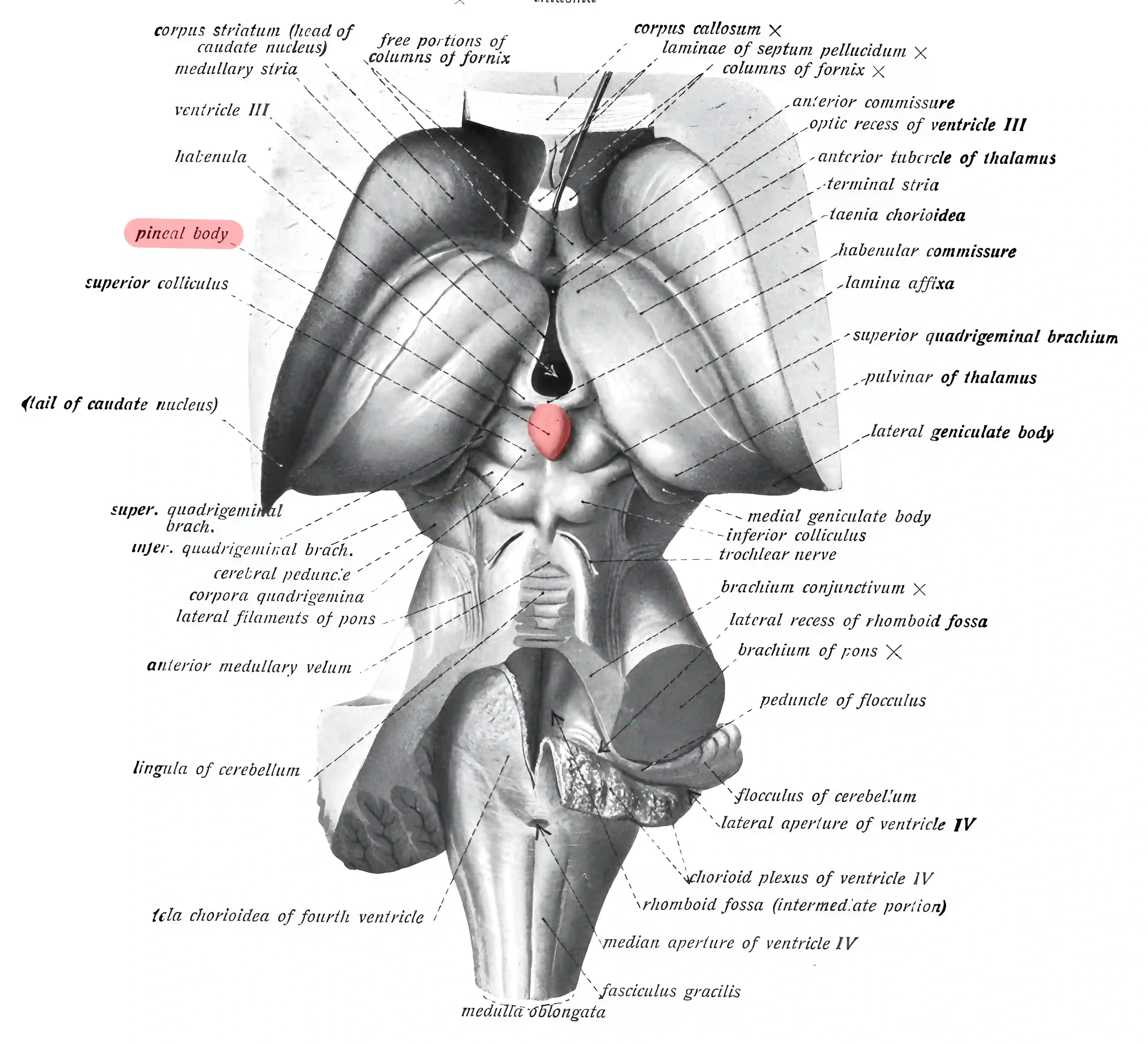
Anatomical drawing of the pineal gland
Schematic depiction of the pineal gland (colored red) on an anatomical drawing from Sobotta's Atlas and Textbook of Human Anatomy, 1908 (Figure 650).
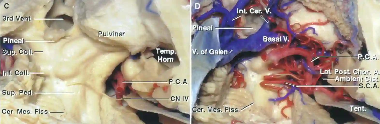
Posterior view of the tectum, Rhoton
Anatomical illustration of the quadrigeminal plate with and without the corresponding vascular structures. Illustration adapted from Albert Rhoton 2002 Neurosurgery.
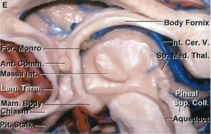
Third ventricle, sagittal view
Sagittal section through the third ventricle. Figure adapted from Albert Rhoton 2002 in Neurosurgery.

Imaging

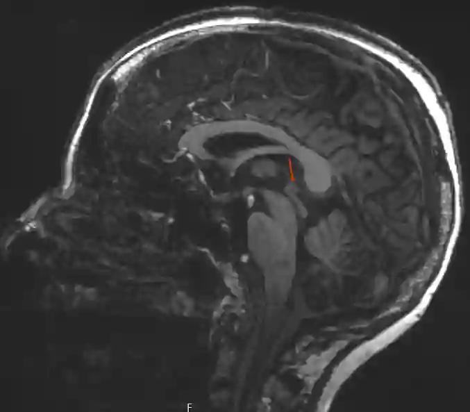
Glandula pinealis on MRI
Visualization of the pineal gland on a sagittal T1-weighted sequence from a 7 Tesla MRI.

Article information

Article created by
Last edited:
on
Revision history
Show revision history (3)

3 edits by 1 author

Edit · Alaric Steinmetz · (+133 −0)

No content change recorded

Edit · Alaric Steinmetz · (+0 −131)

No content change recorded

Article created on · Alaric Steinmetz

Author list:
Newsletter