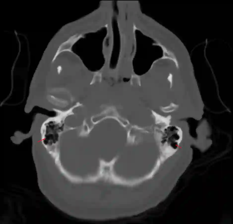
The mastoid cells are pneumatic cavities in the mastoid process of the temporal bone.
Anatomy
The mastoid cells are connected to the tympanic cavity of the middle ear.
Imaging
Mastoid cells can be optimally visualized in CT imaging.

Clinical Relevance
An intraoperative opening of the mastoid cells, such as that which may occur during a retrosigmoid craniotomy, should be sealed cautiously to avoid a cerebrospinal fluid fistula.
Sealing of Intraoperatively Opened Mastoid Cells
Intraoperatively opened mastoid cells can, for example, be sealed by applying a combination of bone wax, muscle, and fibrin glue.