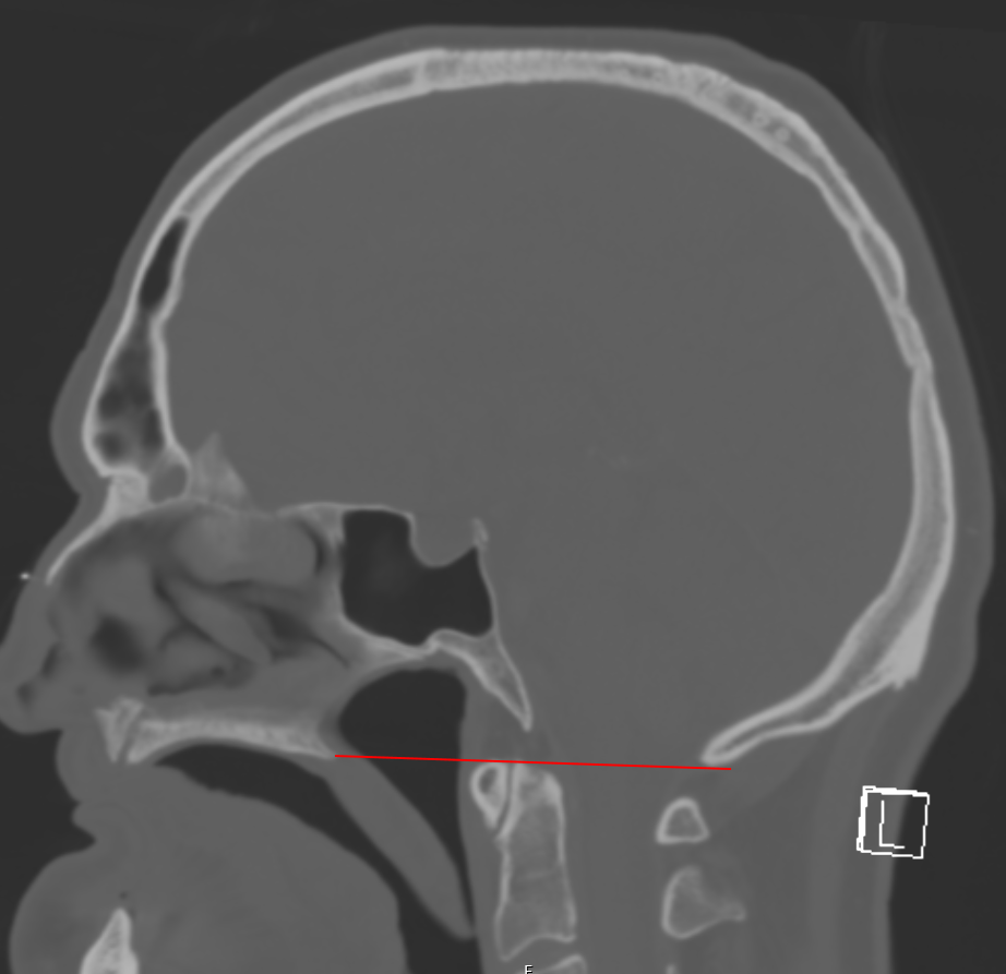
La línea de McGregor conecta el borde posterior del paladar duro con el punto más caudal del occipital1.
Interpretación
La apófisis odontoides del axis no debería sobrepasar esta línea en más de 4,5 mm2. En las imágenes de MRI3 y CT4, la apófisis odontoides del axis se encuentra, en promedio, 0,8 (± 2.4) mm por encima de esta línea.
Diagnóstico por imagen
Referencias
McGregor J. The Significance of Certain Measurements of the Skull in the Diagnosis of Basilar Impression. Br J Radiol. 1948;21:171-181 ↩ ↩
Imaging and Diagnostics. In: Greenberg M, ed. Handbook of Neurosurgery. 9th Edition. Thieme; 2019. ↩ ↩
Cronin CG, Lohan DG, Mhuircheartigh J N, et al. MRI evaluation and measurement of the normal odontoid peg position. Clin Radiol. 2007;62:897-903. ↩ ↩
Cronin CG, Lohan DG, Mhuircheartigh J N, et al. CT evaluation of Chamberlains, McGregors, and McRaes skull-base lines. Clin Radiol. 2009;64:64-69. ↩ ↩